
Breast Calcifications
Calcifications are small calcium deposits that can occur anywhere in the breast tissue. They appear as small white dots that are better seen on mammogram. They are rarely seen on ultrasound and never show up on breast MRIs.
Calcifications are common presentation post menopausal and elderly women as aging can result in benign cells to develop into calcifications or hardening of the breast tissues
Common benign causes of calcifications on MMGs are
- Long standing benign growths in the breast eg Fibroadenoma
- Calcium deposits or buid-ups in the breast blood vessels. Eg atherosclerosis
- Previous injuries or breast infections.
- Past radiation therapy to the breasts
In some cases, calcifications can be an indicator of underlying breast cancer development and can be associated with Ductal Carcinoma in Situ (DCIS) or Invasive Ductal Carcinoma (IDC)
Certain features/ patterns of calcifications on MMG will indicate if the calcifications are suspicious or not.
Benign
Large calcifications / Macrocalcifications, well-defined edges and shapes and spread out.
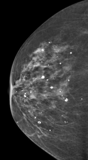
Suspicious
Small speck / dust like / micro-calcifications, various shapes and sizes, clustered and lined along the duct
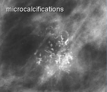
Assessment for suspicious microcalcifications (Birads 4 & 5)
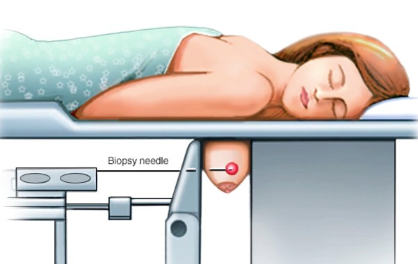
Stereotactic Biopsy
As the calcifications are only seen from MMG; under mammography, the core need biopsy is inserted and biopsy tissue is taken for sampling. The limitation of this technique is inadequate sample of tissue for definitely diagnosis
Sometimes, stereotactic vacuum assisted breast biopsy (VABB) can be offered to remove most of the tissue calcifications to yield a better result compared to stereotactic core biopsy sampling.
credit source : mayo clinic research
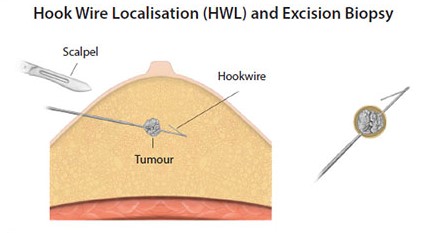
Stereotactic Hook Wire Localization and Excision Biopsy
If the area or pattern of microcalcifications are wider or location is more challenging for core biopsy, then a hook wire is inserted under mammography to help localize the tissue. Once the hook wire is in place within the calcifications, the patient is sent to the operating room for the surgical excision biopsy of the breast lesions.
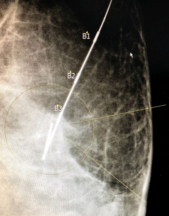
The hook wire in position showing the calcifications adjacent to the marker B1
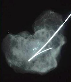
Specimen radiograph after breast tissue removed with hook wire inplace showing the calcifications are included in the tissue